Mycobacterium phage Integral

Add or modify phage thumbnail images to appear at the top of this page.
Know something about this phage that we don't? Modify its data.
| Detailed Information for Phage Integral | |
| Discovery Information | |
| Isolation Host | Mycobacterium smegmatis mc²155 |
| Found By | Lauren Braley, Tara Goble |
| Year Found | 2017 |
| Location Found | Pullman, WA USA |
| Finding Institution | Washington State University |
| Program | Science Education Alliance-Phage Hunters Advancing Genomics and Evolutionary Science |
| From enriched soil sample? | Yes |
| Isolation Temperature | Not entered |
| GPS Coordinates | Unavailable |
| Discovery Notes | The soil sample was collected outside the Shock Physics Building on Washington State University’s campus (Pullman, WA) and across from a construction site. There was evidence of foot, dog, and vehicle traffic with footprint depressions and tire tracks. The temperature reading at the time of soil collection was -1 ̊C, with the collection site being 1x1 cm in area and 0.5 inches in depth. Shrubbery and trees surrounded the area and while no direct water source was visible, the soil was wet. |
| Naming Notes | Integral as in the mathematic term. Also, the word integral is defined as essential or fundamental, much like this bacteriophage's discovery is to the Phage Hunter research. |
| Sequencing Information | |
| Sequencing Complete? | No |
| Genome length (bp) | Unknown |
| Character of genome ends | Unknown |
| Fasta file available? | No |
| Characterization | |
| Cluster | Unclustered |
| Subcluster | -- |
| Annotating Institution | Unknown or unassigned |
| Annotation Status | Not sequenced |
| Plaque Notes | Plaques for the first streak test were pulled from a plaque assay. For the first week of streak plates, as denoted in Figure 2A, the sample pulled showed positive for bacteriophage specific to M. smegmatis. As expected, these plaques were prevalent. A second set of streak tests were then performed from the plaques on plate ‘3G’ (pictured in Figure 2A). From this second set, of the three streak plates, two showed definite plaques while the third was muddled and bacterial growth was more prevalent. It is worthy to note that the bacterial host was having problems in this particular set of streak plates which is evident in the muddled appearance of one of the plates. The pictured plate in Figure 2B was the clearest and was used for the next set of streak tests. Here it is visible to see that the plaques did appear closer to the designated ‘X’ mark. For the final set of purification, plaques came back more defined and clear around the edges. This was the case for some plaques on the plate, but as can be seen in Figure 2C, many of them are also indistinguishable from each other. This, along with the drastic size difference in diameter of the plaques (denoted in Table 1, from 3-4mm to 8-9mm), indicates that this bacteriophage is aggressive. There were some plaques that samples were pulled from, but most were pressed together and most of the bacterial host had been consumed. With the samples collected, the next step from these results was to perform a high titer lysate. |
| Has been Phamerated? | No |
| Publication Info | |
| Uploaded to GenBank? | No |
| GenBank Accession | None yet |
| Refseq Number | None yet |
| Archiving Info | |
| Archiving status | Archived |
| Pitt Freezer Box# | 63 |
| Pitt Freezer Box Grid# | A7 |
| Available Files | |
| Plaque Picture | Download |
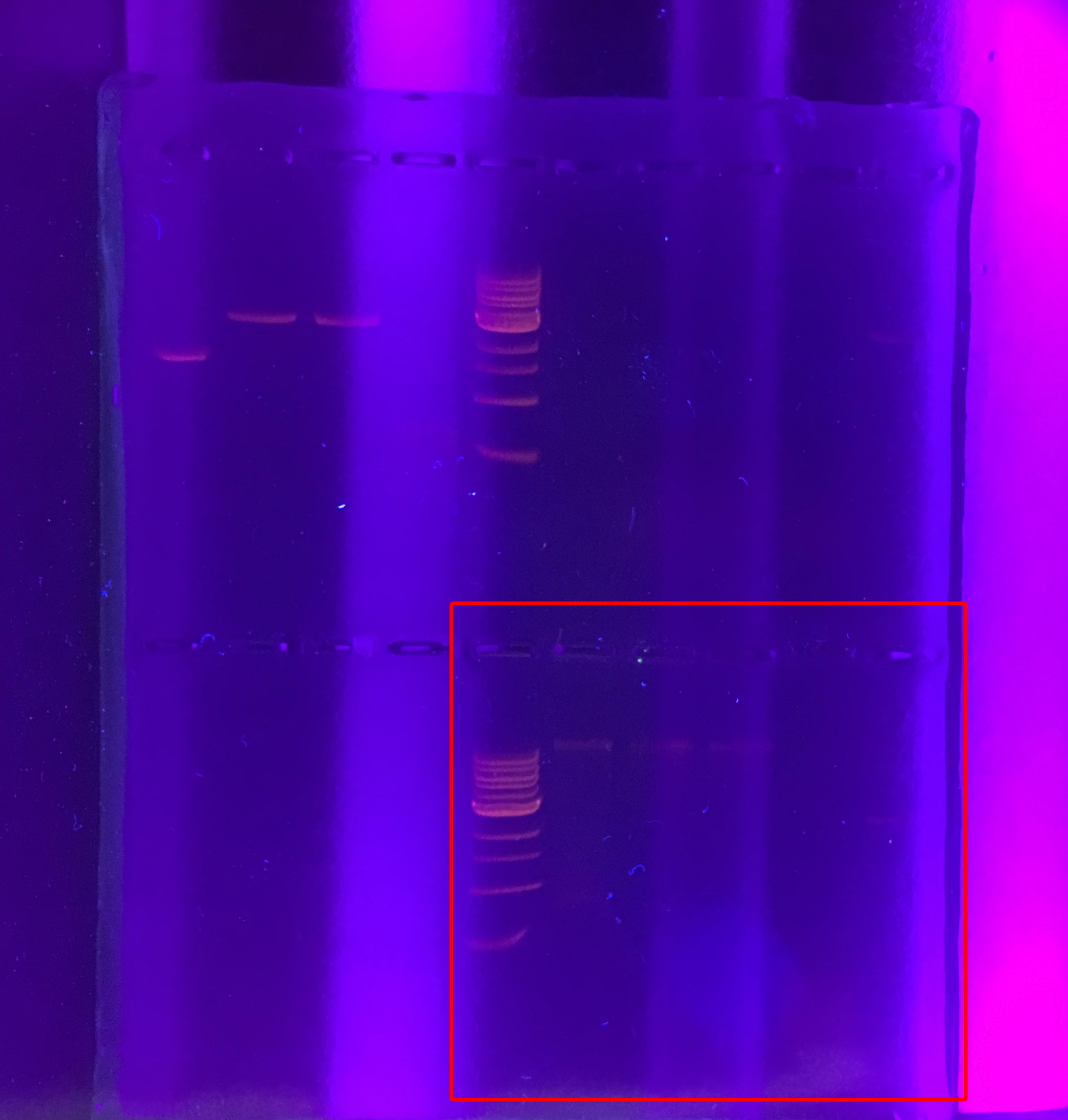
| Restriction Digest Picture | Download |