Mycobacterium phage Genesis
Know something about this phage that we don't? Modify its data.
| Detailed Information for Phage Genesis | |
| Discovery Information | |
| Isolation Host | Mycobacterium smegmatis mc²155 |
| Found By | Justin Yeh |
| Year Found | 2012 |
| Location Found | Baltimore, MD USA |
| Finding Institution | Johns Hopkins University |
| Program | Science Education Alliance-Phage Hunters Advancing Genomics and Evolutionary Science |
| From enriched soil sample? | Yes |
| Isolation Temperature | Not entered |
| GPS Coordinates | 39.326147 N, 76.616942 W Map |
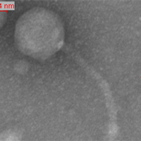
| Discovery Notes | My soil sample that contained the phage was obtained from the Johns Hopkins University Community Garden. Enrichment was done to the sample. My lysates and DNA are highly concentrated. The phage (on EM) is average sized: Head = 80 nm Tail = 165 nm |
| Naming Notes | JHU Phage Hunting gave me the opportunity to apply what I learned in my high school biology courses to real life. Using all the equipment I had read about (but never seen), and performing all the tedious procedures, I finally isolated a phage. This process however, was not smooth and easy-and I was humbled by all the mistakes I made and fascinated by all the progressions. After I saw my phage, my classmates' phages, and their subsequent shouts of fascination pent up from the struggles beforehand, I knew that the genesis of our career as full-time biology students had begun. And I named my phage Genesis. |
| Sequencing Information | |
| Sequencing Complete? | No |
| Genome length (bp) | Unknown |
| Character of genome ends | Unknown |
| Fasta file available? | No |
| Characterization | |
| Cluster | F |
| Subcluster | F1 |
| Cluster Life Cycle | Temperate |
| Lysogeny Notes | Genesis is a Lysogenic Phage. |
| Annotating Institution | Unknown or unassigned |
| Annotation Status | Not sequenced |
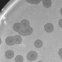
| Plaque Notes | My plaques are large, cloudy, circular plaques with a bull's eye in the center. Like most large plaques, they tend to shrink in size when they are crowded together in plates that are plated with more concentrated dilutions (i.e.: web plates). They are on average about 8.5 mm across (diameter). On more concentrated dilution plates, the plaques shrink down to 7.0-7.5 mm across. The plaques still display the bull's eye even when they shrink in size. |
| Morphotype | Siphoviridae |
| Has been Phamerated? | No |
| Publication Info | |
| Uploaded to GenBank? | No |
| GenBank Accession | None yet |
| Refseq Number | None yet |
| Archiving Info | |
| Archiving status | Archived |
| SEA Lysate Titer | 3.6x10^10 pfu/mL |
| Date of SEA Lysate Titering | Dec 3, 2012 |
| Pitt Freezer Box# | 13 |
| Pitt Freezer Box Grid# | B4 |
| Available Files | |
| Plaque Picture | Download |
| EM Picture | Download |

