Mycobacterium phage Shworlde
Add or modify phage thumbnail images to appear at the top of this page.
Know something about this phage that we don't? Modify its data.
| Detailed Information for Phage Shworlde | |
| Discovery Information | |
| Isolation Host | Mycobacterium smegmatis mc²155 |
| Found By | Julia Holt and Bella Romano |
| Year Found | 2023 |
| Location Found | North Andover, MA United States of America |
| Finding Institution | Merrimack College |
| Program | Science Education Alliance-Phage Hunters Advancing Genomics and Evolutionary Science |
| From enriched soil sample? | Yes |
| Isolation Temperature | 37°C |
| GPS Coordinates | 42.66634 N, 71.12236 W Map |
| Discovery Notes | This sample, sample number 1, was taken at 8:54 am behind the Ash Center at Merrimack college. The coordinates of the location were 42.66634 N and 71.12236 W. There had been rainfall the night before (approximately 1.2”) and the dirt appeared to be partially run off. The sample was collected directly from the surface as everything was damp due to rain. Gloves were worn and a bag was flipped inside out to grab the clump of dirt using the bag. The bag that was used to grab the dirt was placed inside another sandwich bag before being placed inside of a fridge. The sample was collected on 9/12/23 and the weather was 69 degrees Fahrenheit (20.5 degrees Celsius) there was 95% humidity and a 3 mph wind. |
| Naming Notes | Named after a slang term for word for gen z. |
| Sequencing Information | |
| Sequencing Complete? | No |
| Genome length (bp) | Unknown |
| Character of genome ends | Unknown |
| Fasta file available? | No |
| Characterization | |
| Cluster | Unclustered |
| Subcluster | -- |
| Annotating Institution | Unknown or unassigned |
| Annotation Status | Not sequenced |
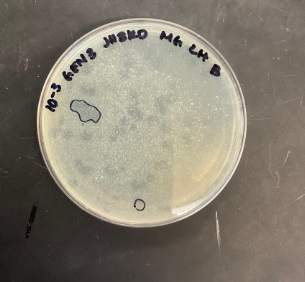
| Plaque Notes | The phage picked from these plates, which was used to form generation two, was picked from the 10^-3 plate. This phage measured approximately .21 cm and was a cloudy plaque which contained cloudy edges which symbolizes it was most likely lysogenic. There were more than 16 plaques that could be distinguished from the larger plaque which takes up the majority of the plate. The plate appeared heterogeneous as there were some plaques which appeared to be more clear than others. The other plates also showed plaques with similar qualities, but the plaques on the 10^-5 plate seemed to have slightly more defined edges. There are less and less phages present as the concentration goes down. The most phages are present in concentration 10^-1 and the least are present in the concentration of 10^-5. This was the second time that we did generation two as the first time 10 microliters of phage were added to the plate and it wiped out the bacteria. Figure 3- image of the three plates which contain dilutions of the phages at 10^-1, 10^-3, and 10^-5 from left to right. All plates in this figure are from generation 1. Generation 2- After 48 hours the three plates were observed. Once again a plaque was picked from the dilution of 10^-3. This phage measured approximately .3 cm in diameter and similarly to the other phage this phage was cloudy, and it had cloudy edges. This plate had more defined plaques, and they appeared overall smaller than those from the last generation. There were over 30 plaques from this generation. Plaques were picked from the 10^-3 plate as they were more defined/separated than those on the 10^-1. The plaques on the 10^-3 plate were also far more visible than those on the 10^-5 plate. This was the second time that we did generation two |
| Has been Phamerated? | No |
| Publication Info | |
| Uploaded to GenBank? | No |
| GenBank Accession | None yet |
| Refseq Number | None yet |
| Archiving Info | |
| Archiving status | Archived |
| Pitt Freezer Box# | 182 |
| Pitt Freezer Box Grid# | C9 |
| Available Files | |
| Plaque Picture | Download |
| Restriction Digest Picture | Download |