Mycobacterium phage JayRo
Know something about this phage that we don't? Modify its data.
| Detailed Information for Phage JayRo | |
| Discovery Information | |
| Isolation Host | Mycobacterium smegmatis mc²155 |
| Found By | Jonathan B. Juste, Ramiro Cisneros |
| Year Found | 2017 |
| Location Found | Las Vegas, NV USA |
| Finding Institution | University of Nevada Las Vegas |
| Program | Science Education Alliance-Phage Hunters Advancing Genomics and Evolutionary Science |
| From enriched soil sample? | Yes |
| Isolation Temperature | 34°C |
| GPS Coordinates | 36.233013 N, 115.316523 W Map |
| Discovery Notes | Soil sample was collected on 09/04/2017, during the evening at approximately 6:00 pm. The ambient temperature for the time of collection was 34.4 °C. It was shady day and the sample was collected from a patch of dry soil. The soil had organic compost added three months prior; it consisted of sandy, rocky dirt with dried leaves interspersed throughout. The soil sample was extracted at a depth of approximately six inches at the base of a pomegranate tree. |
| Naming Notes | The phage name is derived from a combination of the names of the two students who first isolated this virus. JayRo is a manifestation of the first initial of the name "Jonathan" and the last syllable of the name "Ramiro". |
| Sequencing Information | |
| Sequencing Complete? | No |
| Genome length (bp) | Unknown |
| Character of genome ends | Unknown |
| Fasta file available? | No |
| Characterization | |
| Cluster | Unclustered |
| Subcluster | -- |
| Annotating Institution | Unknown or unassigned |
| Annotation Status | Not sequenced |
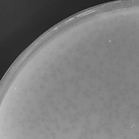
| Plaque Notes | The JayRo isolate plaque morphology is characteristically very small and turbid. Plaques are approximately 0.2 mm in diameter. The edges of the plaques are very smooth and the plaques are almost exactly circular in shape. The margins between plaques are generally even and best observed in plates with a serial dultion factor of 10^-1 or 10^-2. The plaques formed are very consistent in terms of size and appearance through several rounds of purification and amplification on growth media. For ideal observation and examination, plates with plaques should be illuminated with a strong light source since the plaques are quite turbid and difficult to see with the unassisted eye. |
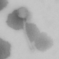
| Morphotype | Siphoviridae |
| Has been Phamerated? | No |
| Publication Info | |
| Uploaded to GenBank? | No |
| GenBank Accession | None yet |
| Refseq Number | None yet |
| Archiving Info | |
| Archiving status | Archived |
| SEA Lysate Titer | 8.3 X 10^9 PFU/mL |
| Pitt Freezer Box# | 52 |
| Pitt Freezer Box Grid# | C12 |
| Available Files | |
| Plaque Picture | Download |
| Restriction Digest Picture | Download |
| EM Picture | Download |

