Mycobacterium phage Lexipuff
Know something about this phage that we don't? Modify its data.
| Detailed Information for Phage Lexipuff | |
| Discovery Information | |
| Isolation Host | Mycobacterium smegmatis mc²155 |
| Found By | Yuki Inaba |
| Year Found | 2013 |
| Location Found | Providence, RI USA |
| Finding Institution | Brown University |
| Program | Science Education Alliance-Phage Hunters Advancing Genomics and Evolutionary Science |
| From enriched soil sample? | Yes |
| Isolation Temperature | Not entered |
| GPS Coordinates | 41.828333 N, 71.401667 W Map |
| Discovery Notes | The soil sample in which I found my phage came from in front of Sidney Frank Hall for Life Sciences near the Pembroke area of Brown University’s campus. It was a nice, sunny day outside with a temperature of around 70°F. I dug about 4.5cm into the soil in order to get a relatively damp soil in which phage are more likely to grow. I filled the conical tube about half full with the soil sample, using a sterile tongue depressor to scoop up the soil. |
| Naming Notes | I named my phage “Lexipuff,” a nickname by which I call one of my closest friends. I dedicated my phage to my friend because my friend makes me so happy, just as discovering my phage brought me happiness and a sense of success. |
| Sequencing Information | |
| Sequencing Complete? | No |
| Genome length (bp) | Unknown |
| Character of genome ends | Unknown |
| Fasta file available? | No |
| Characterization | |
| Cluster | Unclustered |
| Subcluster | -- |
| Annotating Institution | Unknown or unassigned |
| Annotation Status | Not sequenced |
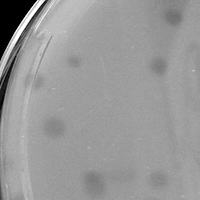
| Plaque Notes | The plaque morphology for Lexipuff was strange in that the phage created two kinds of plaques: smaller ones and bigger ones. The difference in sizes of the plaques was very slight, but I was concerned that I had two phage instead of just one. I purified the phage six times and kept getting the same two kinds of morphologies. I also “picked” one of each kind of plaque and purified them in separate serial dilutions, but I ended up getting the same two kinds of plaques in both serial dilutions. Therefore, I concluded that I only had one phage that was making two different kinds of plaques. Most of the plaques were cloudy instead of clear. |
| Has been Phamerated? | No |
| Publication Info | |
| Uploaded to GenBank? | No |
| GenBank Accession | None yet |
| Refseq Number | None yet |
| Archiving Info | |
| Archiving status | Archived |
| SEA Lysate Titer | 1.7x10^11 |
| Pitt Freezer Box# | 4 |
| Pitt Freezer Box Grid# | D9 |
| Available Files | |
| Plaque Picture | Download |
| Restriction Digest Picture | Download |
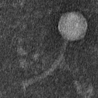
| EM Picture | Download |

