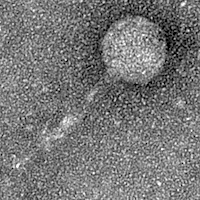
Arthrobacter phage Luna773
Add or modify phage thumbnail images to appear at the top of this page.
Know something about this phage that we don't? Modify its data.
| Detailed Information for Phage Luna773 | |
| Discovery Information | |
| Isolation Host | Arthrobacter globiformis B-2979 |
| Found By | Kulsum Ali |
| Year Found | 2024 |
| Location Found | Bartlett, IL United States |
| Finding Institution | Benedictine University |
| Program | Science Education Alliance-Phage Hunters Advancing Genomics and Evolutionary Science |
| From enriched soil sample? | No |
| Isolation Temperature | 22°C |
| GPS Coordinates | 41.99772 N, 88.24846 W Map |
| Discovery Notes | The soil was moist when it was collected because the snow was melting. When collected at 12:30 PM the entire area was soft. I used a shovel and dug about 1 inch. deep avoiding the rocks to extract the soil. the soil I collected was dark because it was moist and it contained some rocks and grass. |
| Naming Notes | I named my phage after my cat. |
| Sequencing Information | |
| Sequencing Complete? | No |
| Genome length (bp) | Unknown |
| Character of genome ends | Unknown |
| Fasta file available? | No |
| Characterization | |
| Cluster | Unclustered |
| Subcluster | -- |
| Annotating Institution | Unknown or unassigned |
| Annotation Status | Not sequenced |
| Plaque Notes | Once observing these plates it was concluded that there was contamination and many plaques were visible. On plate #1 (right side) which contained 200 uLs (Figure 1) the agar was still smooth, most of the phage plaques were all small and fuzzy and had a diameter of 1 mm and the biggest one was about 3 mm. The number of plaques present in plate #1 was approximately 45 and they were all visible. On plate #2 which contained 400uLs (Figure 1) the agar was more contaminated then #1, it contained a lot of plaques in different size and those that were bigger were more clear instead of cloudy.The plaques were everywhere and visiblemaking it too difficult to count the exact amount. Both plates are potentially lytic. This procedure was then repeated again as Direct Isolation and Plaque assay Part 2. In this procedure more direct f.s was supposed to be added into the bacteria about 300 uLs and 600 uLs. However, due to a misunderstanding between the lab conductor only 250 and 500 uLs were added. This should not make much of difference because plaques were already shown with a small amount of bacteria added. Once conducted, the results were similar to those of the first one. Plate #1 (right side) contained many plaques about 60 andthe plate #2 was fully contaminated with plaques making it difficult to approximate. |
| Has been Phamerated? | No |
| Publication Info | |
| Uploaded to GenBank? | No |
| GenBank Accession | None yet |
| Refseq Number | None yet |
| Archiving Info | |
| Archiving status | Archived |
| Pitt Freezer Box# | 218 |
| Pitt Freezer Box Grid# | F5 |
| Available Files | |
| Plaque Picture | Download |
| EM Picture | Download |
