Mycobacterium phage Saitama
Know something about this phage that we don't? Modify its data.
| Detailed Information for Phage Saitama | |
| Discovery Information | |
| Isolation Host | Mycobacterium smegmatis mc²155 |
| Found By | Anastasia Guseva and Breanna Edinger |
| Year Found | 2016 |
| Location Found | Corpus Christi, TX USA |
| Finding Institution | Del Mar College |
| Program | Science Education Alliance-Phage Hunters Advancing Genomics and Evolutionary Science |
| From enriched soil sample? | Yes |
| Isolation Temperature | 37°C |
| GPS Coordinates | Unavailable |
| Discovery Notes | The soil/medium in which the phage was collected from was extracted on August 29th, 2016 at precisely 12:21 p.m. The area was near a drainage close to a building at the college, however, it was enclosed and nearly completely shaded with the shrubbery and plant-life that adorned the area. It was secluded and quite dark with very little light penetration. The weather at the time was mostly cloudy with a humid-type of air quality. The temperature was hot with a temperature of 28.9 degrees Celsius (84 degrees Fahrenheit). The soil itself was dark and dry with a clay-like texture that clumped together in small solids. |
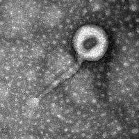
| Naming Notes | The reason the name was chosen was due to the fact that the general physical appearance of the phage EM image were similar to a character my partner and I both knew. Both shared the similarities of a round, almost bald-like shape of a head that was more noticeable than the rest of their forms. They both tend to blend in, though in the case of the phage that is mostly due to lingering debris in the image rather than an official characteristic. |
| Sequencing Information | |
| Sequencing Complete? | No |
| Sequencing Facility | University of North Texas |
| Shotgun Sequencing Method | Sanger |
| Genome length (bp) | Unknown |
| Character of genome ends | Unknown |
| Fasta file available? | No |
| Characterization | |
| Cluster | Unclustered |
| Subcluster | -- |
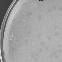
| Lysogeny Notes | The plaques appeared to be lysogenic since the plaques had appeared turbid (not transparent), therefore, the cell hadn't been completely destroyed, a factor that would set it aside as lytic. |
| Annotating Institution | Unknown or unassigned |
| Annotation Status | Not sequenced |
| Plaque Notes | The phage plaques were small with a slightly opaque form. They appear as dots or spots and rarely deviated in size unless clumped together. The small size of the plaques might be due to less phage or simply because of a characteristic of our phage singularly. |
| Morphotype | Siphoviridae |
| Has been Phamerated? | No |
| Publication Info | |
| Uploaded to GenBank? | No |
| GenBank Accession | None yet |
| Refseq Number | None yet |
| Archiving Info | |
| Archiving status | Archived |
| Pitt Freezer Box# | 34 |
| Pitt Freezer Box Grid# | H5 |
| Available Files | |
| Plaque Picture | Download |
| EM Picture | Download |

