Mycobacterium phage Tinkcarose
Add or modify phage thumbnail images to appear at the top of this page.
Know something about this phage that we don't? Modify its data.
| Detailed Information for Phage Tinkcarose | |
| Discovery Information | |
| Isolation Host | Mycobacterium smegmatis mc²155 |
| Found By | Maddi Carini and Olivia Tinkham |
| Year Found | 2023 |
| Location Found | North Andover, MA United States of America |
| Finding Institution | Merrimack College |
| Program | Science Education Alliance-Phage Hunters Advancing Genomics and Evolutionary Science |
| From enriched soil sample? | Yes |
| Isolation Temperature | 37°C |
| GPS Coordinates | 42.402 N, 71.722 W Map |
| Discovery Notes | Sampling was done outside of Deegan West. This sample was taken at 285° W, 42°40’2” N 71°7’22”W North Andover, MA at 230 ft elevation. A pair of gloves were put on to grab the damp soil sample. Once gloves were on, the plastic bag was opening and took a chunk of surface level dirt. The grab for soil did not go beyond the surface layer of the ground. A handful of the damp soil and put it into the plastic sandwich baggy. Onced it was in the baggy it was sealed shut so that nothing could enter the bag and contaminate the soil. The color of the soil on the ground was a light brown color, but once added to the plastic bag it was a darker brown color. This sample was wet but still solid due to the fact it rained the whole day before collecting the sample. This sample was taken on 09/19/2023 at 9:30 AM with the initials OT written on the bag. The temperature for the day that the soil was collected was 63° and the humidity was 91%. |
| Naming Notes | Named after the names of the two students who discovered the phage. |
| Sequencing Information | |
| Sequencing Complete? | No |
| Genome length (bp) | Unknown |
| Character of genome ends | Unknown |
| Fasta file available? | No |
| Characterization | |
| Cluster | Unclustered |
| Subcluster | -- |
| Annotating Institution | Unknown or unassigned |
| Annotation Status | Not sequenced |
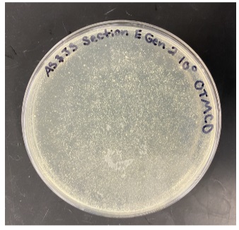
| Plaque Notes | The only image shown is the 100 concentration of plaques. The pictures of the 10-3 concentration and the 10-5 concentration are omitted from the figure because the plates were completely clear and did not have any bacteria on them. This is most likely due to error in the process of handling the bacteria when transferring the contents of the microcentrifuge tubes to the 14mL tubes, causing nothing to show up at all on the plate. The second generation assay was created from a phage within the first generation. The 100 concentration also overall does not display a very concentrated sample, but there are a few noticeable clear plaques at the upper right. The upper right is where we took our isolated plaque from in order to create generation three. The plaques in the 100 concentration were clear, meaning that they were lytic, and there was no evidence of cloudiness in the center of the plaque or around the plaques. |
| Has been Phamerated? | No |
| Publication Info | |
| Uploaded to GenBank? | No |
| GenBank Accession | None yet |
| Refseq Number | None yet |
| Archiving Info | |
| Archiving status | Archived |
| Pitt Freezer Box# | 182 |
| Pitt Freezer Box Grid# | C10 |
| Available Files | |
| Plaque Picture | Download |
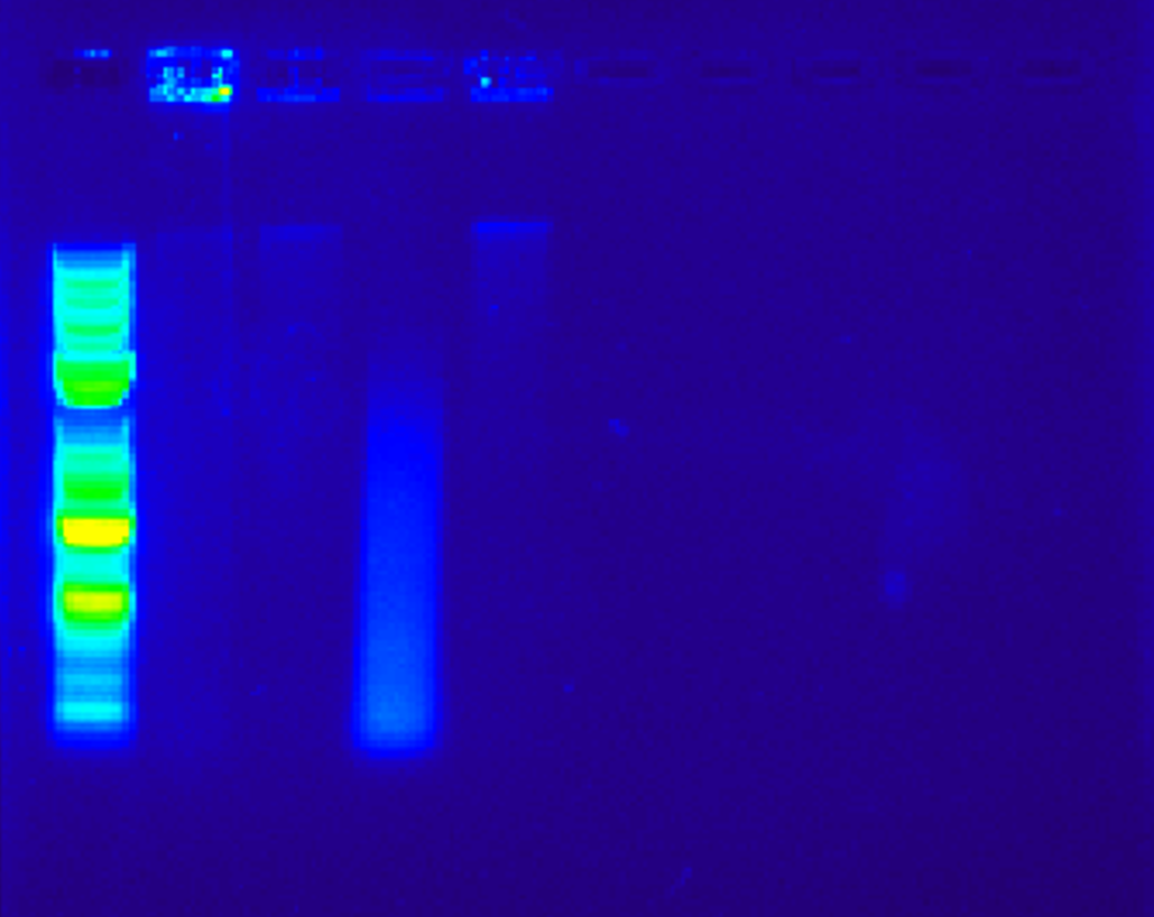
| Restriction Digest Picture | Download |