Gordonia phage Zarambe
Know something about this phage that we don't? Modify its data.
| Detailed Information for Phage Zarambe | |
| Discovery Information | |
| Isolation Host | Gordonia terrae 3612 |
| Found By | Brijai Varma |
| Year Found | 2018 |
| Location Found | Pittsburgh, PA USA |
| Finding Institution | University of Pittsburgh |
| Program | Science Education Alliance-Phage Hunters Advancing Genomics and Evolutionary Science |
| From enriched soil sample? | No |
| Isolation Temperature | 30°C |
| GPS Coordinates | 40.4436 N, 79.9602 W Map |
| Discovery Notes | The condition of the soil was very dry and fine with some crumbled dirt and was a light, washed out brown color. The nearby vegetation is shown in the picture with many bushes and one medium sized tree present in the surrounding area. These pictures were taking on September 3rd, 2018 at 5:13 PM, with sunny clear skies and a temperature of 91 degrees Fahrenheit. The humidity was 46% with winds blowing S.W. at 6 mph. The location's coordinates are 40.4436 for Latitude and -79.9602 for Longitude. The soil sample was was stored in a room temperature drawer at 72 degree Fahrenheit for approximately 16 hours. The sample was then transported to the lab in a span of 3 hours. |
| Sequencing Information | |
| Sequencing Complete? | No |
| Genome length (bp) | Unknown |
| Character of genome ends | Unknown |
| Fasta file available? | No |
| Characterization | |
| Cluster | Unclustered |
| Subcluster | -- |
| Lysogeny Notes | No lysogen produced due to contamination. |
| Annotating Institution | Unknown or unassigned |
| Annotation Status | Not sequenced |
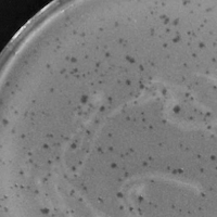
| Plaque Notes | On the plate, the top left side with the opaque yellow white tint describes negative results and contamination from other factors. However, the entire other side of the plate contains multiple large plaques that do not have clear borders. There are many small specs left over of the orange top layer of agar as most of the top layer is cleared. The top layer of agar has a rough texture. For the spot test, there were three, large circular spots on the plate. There were no signs of contamination, as the top layer agar was mostly a smooth surface with orange tint except for the three large circular plaques, which were clear in color and in terms of clarity, were mostly transparent. These descriptions relate to positive results for the spot test. |
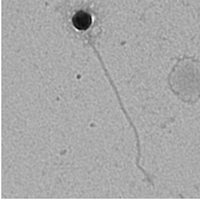
| Morphotype | Siphoviridae |
| Has been Phamerated? | No |
| Publication Info | |
| Uploaded to GenBank? | No |
| GenBank Accession | None yet |
| Refseq Number | None yet |
| Archiving Info | |
| Archiving status | Archived |
| Pitt Freezer Box# | 91 |
| Pitt Freezer Box Grid# | E3 |
| Available Files | |
| Plaque Picture | Download |
| Restriction Digest Picture | Download |
| EM Picture | Download |

