| Detailed Information for Phage Butters | |
| Discovery Information | |
| Isolation Host | Mycobacterium smegmatis mc²155 |
| Found By | Lena Ma |
| Year Found | 2011 |
| Location Found | Philadelphia, PA USA |
| Finding Institution | Lehigh University |
| Program | Science Education Alliance-Phage Hunters Advancing Genomics and Evolutionary Science |
| From enriched soil sample? | Yes |
| Isolation Temperature | Not entered |
| GPS Coordinates | 40.06067 N, 75.046608 W Map |
| Discovery Notes | Phage was discovered from a soil sample in Philadelphia at 25 degrees Celcius. The temperature is sunny and 78% humidity. The soil sample collected was dry and obtained from soil 3cm deep. The soil was also near growing plants where there was only sunlight (no shade). |
| Sequencing Information | |
| Sequencing Complete? | Yes |
| Date Sequencing Completed | Feb 29, 2012 |
| Sequencing Facility | Virginia Commonwealth University Nucleic Acids Research Facilities |
| Shotgun Sequencing Method | 454 |
| Sequencer Used | 454 Genome Sequencer FLX |
| Approximate Shotgun Coverage | 280 |
| Genome length (bp) | 41491 |
| Character of genome ends | 3' Sticky Overhang |
| Overhang Length | 13 bases |
| Overhang Sequence | CCCGCCGCCCTCG |
| GC Content | 65.8% |
| Fasta file available? | Yes: Download fasta file |
| Characterization | |
| Cluster | N |
| Subcluster | -- |
| Cluster Life Cycle | Temperate |
| Other Cluster Members |
Click to ViewAggie Andies BabeRuth Bosection6 Butters Carcharodon Charlie Chewbacca Cubone Duplicity EGUnicorn Fulbright Gex Hanako Impisi Jamie19 Journey Kevin1 Magsby Melville MichelleMyBell Nenae Panchino Parmesanjohn PhancyPhin Philonius Phloss Phrann Pipsqueaks Purgamenstris Raymond7 Rebel Redi Rubeelu Schnauzer Scitech ShrimpFriedEgg Shweta Silvafighter Silvy SkinnyPete Smurph Snekmaggedon Spinach SpongeBob Tapioca Tortoise12 Xeno Xerxes |
| Annotating Institution | Unknown or unassigned |
| Annotation Status | In GenBank |
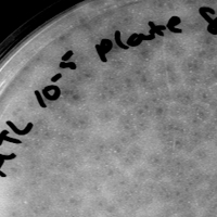
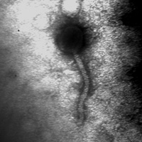
| Plaque Notes | After 24 hours of incubation, the plaque sizes are all 0.5mm in diameter and turbid. After 48 hours of incubation the plaques grew to be two different sizes. The first size is 0.5mm-1.0mm in diameter and turbid. The second size is 2.0mm-3.0mm in diameter and turbid. Even with the two different sizes the plaques yield the same phage. After incubation for longer than 48 hours, the plaques are all 3.0mm in diameter and bullseye. The bullseye effect is only shown after incubation of longer than 48 hours. Titers were performed from one of the 0.5mm-1.0mm plaques and from one of the 2.0mm-3.0mm plaques and both titers yielded the same morphology as discussed above, thus concluding that even with the two sized plaques, the phage is the same. The phage is a siphoviridae with an icosahedral head but looks almost spherical on the EM picture. It has a 3:1 tail:head ratio and has horizontal banding on its tail. There is also a small ball on the end of the tail. |
| Morphotype | Siphoviridae |
| Number of Genes | 66 |
| Number of tRNAs | 0 |
| Number of tmRNAs | 0 |
| Has been Phamerated? | Yes |
| Gene List |
Click to ViewButters_1 Butters_2 Butters_3 Butters_4 Butters_5 Butters_6 Butters_7 Butters_8 Butters_9 Butters_10 Butters_11 Butters_12 Butters_13 Butters_14 Butters_15 Butters_16 Butters_17 Butters_18 Butters_19 Butters_20 Butters_21 Butters_22 Butters_23 Butters_24 Butters_25 Butters_26 Butters_27 Butters_28 Butters_29 Butters_30 Butters_31 Butters_32 Butters_33 Butters_34 Butters_35 Butters_36 Butters_37 Butters_38 Butters_39 Butters_40 Butters_41 Butters_42 Butters_43 Butters_44 Butters_45 Butters_46 Butters_47 Butters_48 Butters_49 Butters_50 Butters_51 Butters_52 Butters_53 Butters_54 Butters_55 Butters_56 Butters_57 Butters_58 Butters_59 Butters_60 Butters_61 Butters_62 Butters_63 Butters_64 Butters_65 Butters_66 |
| Publication Info | |
| Uploaded to GenBank? | Yes |
| GenBank Accession | KC576783 |
| Refseq Number | NC_021061 |
| Published in Paper | Hatfull et al 2013, Genome Announcements: Complete genome sequences of 63 mycobacteriophages |
| Archiving Info | |
| Archiving status | Archived |
| SEA Designator | 2011LEHIbuttersLM |
| Pitt Freezer Box# | 25 |
| Pitt Freezer Box Grid# | 56 |
| Available Files | |
| Plaque Picture | Download |
| Restriction Digest Picture | Download |
| EM Picture | Download |
| Final DNAMaster File | Download |

